This is an education resource only. Ordering of all procedure codes on this website are subject to the Canberra Health Services guidelines for imaging orders.
CT FOOT CONTRAST
FOR ALL EXTREMITIES, PLEASE REFERENCE SIDE AND REGION OF INTEREST TO BE SCANNED IN CT REQUEST

INDICATIONS (1-4)
-
Post-Operative evaluation
-
Foot Soft tissue mass (X-ray and US non-diagnostic & MRI Contraindicated)
-
Osteomyelitis/Septic arthritis of the foot:
-
- Arthroplasty or implanted intra-articular surgical hardware
-
- X-ray suggests effusion or soft tissue swelling
-
Soft tissue infection of the foot:
-
- wound, possible foreign body,
-
- X-ray normal, necrotizing fasciitis highly suspected
-
- Soft tissue gas on radiography, no puncture wound
PATIENT PREPARATION
-
Patient able to lie still for ten minutes
-
Not claustrophobic (sedation may be given)
-
Cognitively capable of following basic instructions
-
Metal artefacts removed from the region of interest
-
No respiratory distress when lying supine
-
Not allergic to Iodine based contrast
-
No known kidney disease (eGFR below 30 as per RANZCR), however, acute setting consultant may sign to continue with poor renal function
-
No hyperthyroidism, may induce thyroid storm
-
Patient to have 20G cannula in anterior cubital fossa.
-
Preferably patient fasted for 4 hours
ANATOMY INCLUDED
SCAN RANGE: Above ankle joint to toes
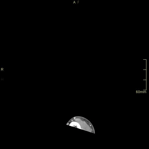
CT Foot Contrast- Soft tissue window (axial)

CT Foot Contrast- Bone window (axial)
CT Foot Contrast- Soft tissue window (coronal)

CT Foot Contrast- Bone window (coronal)

CT Foot Contrast- Soft tissue window (sagittal)

CT Foot Contrast- Bone window (sagittal)
CT FOOT NON CONTRAST
FOR ALL EXTREMITIES, PLEASE REFERENCE SIDE AND REGION OF INTEREST TO BE SCANNED IN CT REQUEST
INDICATIONS (1-4)
-
Chronic Foot pain, gout or pseudogout suspected (x-ray inconclusive), consider dual energy CT if available
-
Primary bone tumour suspected of the Foot- osteoid osteoma suspected on x-ray or clinical exam
-
Soft tissue infection of the foot:
- Necrotizing fasciitis highly suspected
- Soft tissue gas on radiography, no puncture wound -
Foot trauma:
- Dislocation suspected (x-ray non-diagnostic)
- Lis franc injury
- Occult fracture suspected (midfoot instability present, or x-ray non diagnostic) -
Chronic Midfoot pain
- X-ray negative, accessory ossicles suspected
- X-ray negative, occult fracture suspected -
Fracture union and healing
-
Post-operative evaluation
-
Pre-operative planning
PATIENT PREPARATION
-
Patient able to lie still for five minutes
-
Not claustrophobic (sedation may be given)
-
Cognitively capable of following basic instructions
-
Metal artefacts removed from the region of interest
-
No respiratory distress when lying supine
ANATOMY INCLUDED
SCAN RANGE: Above ankle joint to toes

CT Foot Non Contrast- Bone window (axial)
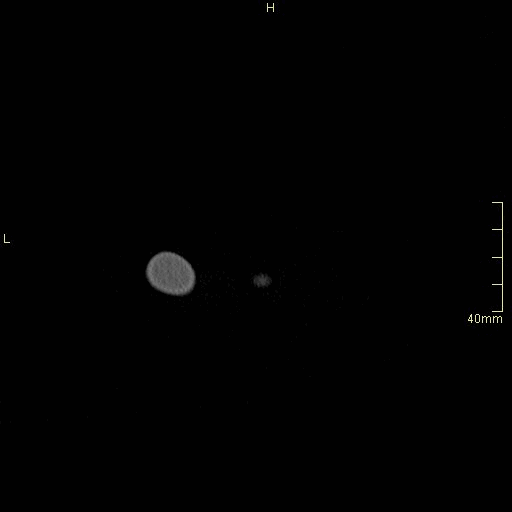
CT Foot Non Contrast- Bone window (coronal)

CT Foot Non Contrast- Bone window (sagittal)

CT Foot Non Contrast- Soft tissue window (axial)
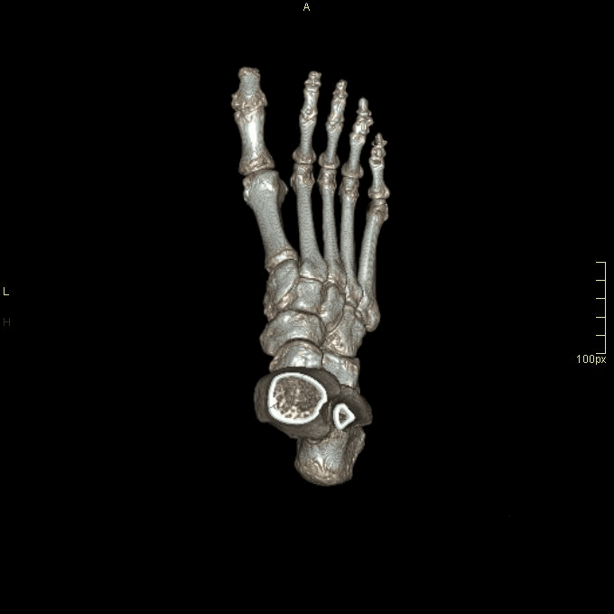

Extra reconstruction: 3D VR
REFERENCES
-
Radiopaedia. CT foot (protocol). [Internet]. 2010 [updated 23 March 2023, cited 04 Feb 2024]. Available from https://radiopaedia.org/articles/ct-foot-protocol-1?lang=gb
-
Melenevsky Y, Mackey R, Abrahams R, Thomson N. Talar Fractures and Dislocations: A Radiologist’s Guide to Timely Diagnosis and Classification. Radiographics. 2015;35(3):765-79. doi:10.1148/rg.2015140156 – Pubmed
-
Johnson, P. T., Fayad, L. M., & Fishman, E. K. (2007). CT of the foot. Journal of Computer Assisted Tomography, 31(6), 961–969. https://doi.org/10.1097/rct.0b013e318043a1d8
-
American College of Radiology (ACR). Appropriateness Criteria. [Internet]. 2022 [Updated 2023, Cited 20 Jan 2024]. Available from https://www.acr.org/Clinical-Resources/ACR-Appropriateness-Criteria